Atopic dermatitis (AD), also known as atopic eczema, is a long-term type of inflammation of the skin. Atopic dermatitis is also often called simply eczema but the same term is also used to refer to dermatitis, the larger group of skin conditions. Atopic dermatitis results in itchy, red, swollen, and cracked skin. Clear fluid may come from the affected areas, which can thicken over time.
Atopic dermatitis affects about 20% of people at some point in their lives. It is more common in younger children. Females are affected slightly more often than males. Many people outgrow the condition.
Scratching the affected areas worsens the eczema and increases the risk of skin infections. Many people with atopic dermatitis develop hay fever or asthma.
Symptoms
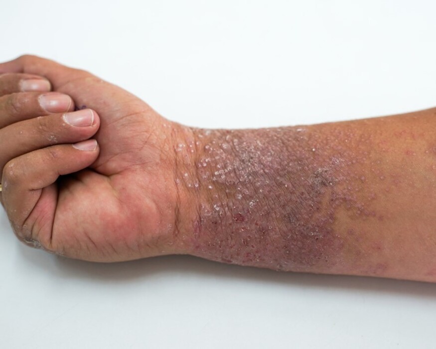
Atopic dermatitis symptoms include intense itching, redness, and dry/scaly skin, which can lead to red, bumpy, or thickened rashes. The rash can also be raw, oozing, or crusted, and may appear in different locations depending on age, such as the cheeks and scalp in infants or the creases of the elbows and knees in children and adults. Skin can darken or lighten in affected areas, and in severe cases, blisters may form.
Itching (Pruritus): This is the primary symptom, often described as “the itch that rashes,” and can be severe.
Redness and Inflammation: Patches of skin appear red and inflamed, which can be particularly noticeable on lighter skin tones.
Dry, Scaly Skin: The skin often feels dry and may become flaky or scaly.
Swelling (oedema).
Scaling cracking (skin fissures).
Rash: A rash is a common sign, which can vary in appearance.
Bumpiness (papulation)
Oozing and Crusting: The rash may leak clear fluid, or it can develop into crusted, weepy sores or blisters, especially if scratched.
Oozing of clear fluid.
Skin Discoloration: In people with darker skin tones, areas of inflamed skin may appear lighter or darker.
Darkening of the skin around the eyes.
Skin Thickening (Lichenification): Repeated scratching and rubbing can cause the skin to become thick, leathery, and more prone to permanent itching.
Raw, Open Sores: Scratching can break the skin, leading to raw, open areas;
Cause
Atopic dermatitis is caused by a weakened skin barrier and an overactive immune system, often due to genetic factors, leading to inflammation and itching when exposed to triggers like allergens (pollen, dust mites, pet dander), irritants (soaps, detergents, rough fabrics), certain foods (eggs, milk, peanuts), environmental factors (dry/cold weather, heat, sweat), skin infections, and stress.
Underlying Causes
Genetics and Immune System:
A genetic tendency for “atopy” (a predisposition to allergic diseases like asthma and hay fever) often underlies atopic dermatitis.
Skin Barrier Dysfunction:
A genetic variant can prevent the skin from forming a strong protective barrier, allowing allergens and irritants to penetrate and trigger inflammation.
Triggers that Worsen Symptoms
Irritants:
Soaps, detergents, shampoos, and harsh chemicals can dry out the skin and cause a flare-up.
Allergens:
Environmental: Dust mites, pet dander, molds, and pollens can trigger symptoms.
Food: Allergies to foods like eggs, peanuts, milk, soy, or wheat can be a trigger, especially in young children.
Environmental Factors:
Climate: Dry, cold weather can lead to dry skin and worsen eczema. Heat, humidity, and sweating can also be triggers.
Pollution: Exposure to tobacco smoke and other pollutants can aggravate symptoms.
Fabrics:
Wearing rough materials like wool or certain synthetic fabrics next to the skin can cause irritation.
Skin Infections:
Bacterial infections, such as those caused by Staphylococcus aureus, are common in atopic dermatitis and can worsen the condition.
Stress:
Psychological stress is a significant factor that can worsen itching and flare-ups.
Hormonal Changes:
Some women may experience worsening symptoms before their period or during pregnancy.
Dry Skin:
Inherently dry skin is a common factor that can make skin more susceptible to irritation and flares.
Diagnosis and Tests
In some cases, your provider may recommend a skin biopsy.
DIAGNOSIS
Atopic dermatitis is diagnosed clinically by a doctor after a physical exam and review of your symptoms and medical history, including personal and family history of allergies or asthma. The diagnosis requires the presence of major features like intense itchiness (pruritus), characteristic skin lesions, a relapsing course, and a history of atopy. In some cases, a skin biopsy may be done to examine a skin sample under a microscope, or allergy tests may be recommended to identify triggers.
Diagnostic Steps
- Physical Examination:
A healthcare provider will examine your skin for typical signs of atopic dermatitis, such as dryness (xerosis), redness, and scaly or thickened skin. - Medical History:
They will ask about your symptoms, when they started, and if you have a personal or family history of allergies, asthma, or other atopic conditions. - Major Diagnostic Criteria:
The diagnosis often relies on meeting certain criteria, such as the presence of pruritus, characteristic lesion appearance and location, chronic relapsing course, and a history of atopy, according to models like the Hanifin-Rajka criteria. - Skin Biopsy (Less Common):
If the diagnosis isn’t clear from the physical exam, a small piece of skin may be removed and examined under a microscope by a dermatopathologist to rule out other skin conditions. - Allergy Testing (Sometimes):
Allergy tests, such as checking IgE levels in the blood, might be recommended to help identify potential environmental or food triggers, though IgE levels are not definitive for atopic dermatitis alone.
Doctor Looks For
Pruritus (Itching):
Xerosis:
Skin Lesions: Redness (erythema), swelling (edema), erosions, crusting, and changes in skin texture called lichenification can be present.
Personal/Family History: A personal or family history of atopy (eczema, asthma, hay fever) is a significant factor.
Age of Onset: Symptoms often appear in infancy or childhood, with a majority of cases developing before age five.
TREATMENT
ment focuses on managing flares and repairing the skin barrier through moisturizing, using medicated creams like corticosteroids and calcineurin inhibitors, and maintaining a consistent, gentle skincare routine. For more severe cases, systemic treatments such as oral medications, biologics, or phototherapy may be necessary. Identifying and avoiding triggers, wearing comfortable clothing, and using antipruritics can also help manage symptoms.
Skincare and Supportive Care
Moisturize regularly:
Apply liberal amounts of a fragrance-free emollient or moisturizer (eg, white petrolatum)
to the skin immediately after bathing to help restore the skin barrier.
Gentle bathing:
Use lukewarm, not hot, water and mild, soap-free cleansers.
Avoid triggers:
Identify and avoid factors that can worsen symptoms, such as certain fabrics, irritants, or allergens.
Wear appropriate clothing:
Choose comfortable, loose-fitting clothing made from smooth, natural fibers like cotton.
Medications
1)Moisturizing creams can help restore the skin barrier.
Barrier repair creams containing ceramides may be used.
2)Topical Corticosteroids: Apply a topical corticosteroid twice daily to affected areas until improvement occurs (usually a few days to 2–3 weeks).
3)Infants (or treatment of the face in a patient of any age): Use a low-potency preparation (eg, hydrocortisone 1% or 2.5%).
Newer (second generation) corticosteroids, such as fluticasone propionate and mometasone furoate, are more effective and safer than older ones.
4)Young children (exclusive of the face): Use a low-potency preparation (eg, hydrocortisone 1% or 2.5%) or, if necessary, a mid-potency preparation (eg, triamcinolone 0.025% or 0.1%, fluocinolone 0.025%).
5)Older children and adolescents (exclusive of the face): Use a mid-potency preparation (eg, triamcinolone 0.1%); a high-potency agent (eg, mometasone 0.1%, fluocinonide 0.05%) may be needed for resistant, non-facial areas during a flare.
6)Calcineurin inhibitors applied to the skin decrease inflammation and help prevent flares.
Pimecrolimus and tacrolimus are suitable for treating atopic dermatitis in sensitive sites such as the eyelids, skin folds, and genital areas.
Examples include tacrolimus (Protopic) and pimecrolimus (Elidel).
7)Phosphodieterase-4 (PDE4) Inhibitors:
Phosphodieterase-4 inhibitors, a topical cream, can help with inflammation when the symptoms do not respond to other treatments.
Such as Zoryve (roflumilast), are a class of non-steroidal medications approved for inflammatory skin conditions like plaque psoriasis and atopic dermatitis.
8)CRISABOROLE OINTMENT: an inhibitor of PDE-4, is also effective and safe as a topical treatment for mild-to-moderate AD.
Approved to treat patients 3 months of age or older, crisaborole can be used on many areas of the skin.
Brand name: Eucrisa
9)Antidepressants and antihistamine: Can help relieve itching by blocking the effects of histamine and to control pruritus (itchiness).
Systemic Medications (for moderate-to-severe disease):
10)Conventional oral medications for AD include systemic immunosuppressants, such as ciclosporin,
methotrexate,
azathioprine, and mycophenolate.
11)Janus Kinase (JAK) Inhibitors: Oral pills that block inflammation from within the cells.
These include dupilumab (Dupixent), tralokinumab (Adtralza, Adbry),
abrocitinib (Cibinqo),
baricitinib (Olumiant)
and upadacitinib (Rinvoq).
Dupilumab is a subcutaneous injection administered every 2 weeks.
Lebrikizumab is also approved in the EU for treating moderate-to-severe AD but in the US its approval was declined due to manufacturing issues.
Nemolizumab (Nemluvio) was approved to treat atopic dermatitis in December 2024.
Nemolizumab has been approved by the FDA for the treatment of moderate-to-severe AD in adults and in pediatric patients 12 years of age and older, in conjunction with topical corticosteroids or calcineurin inhibitors (or both).
12)Biologics: Approved biologics injection
include dupilumab (Dupixent), lebrikizumab
and tralokinumab (Adbry). Injectable medications that target specific immune system functions to control symptoms and manage atopic dermatitis.
Other Therapies
13)Phototherapy: Special light treatment that can help control symptoms.
14)Allergen Immunotherapy: Injections or drops under the tongue to reduce allergic reactions.
15)Wet dressings.
16)Counseling.
17)Relaxation, behavior modification and biofeedback.
Diet
The role of vitamin D on atopic dermatitis is not clear, but vitamin D supplementation may improve its symptoms.
There is no clear benefit for pregnant mothers taking omega 3 long-chain polyunsaturated fatty acid (LCPUFA) in preventing the development of AD in their child.